Home » Without Label » Labeled Diagram Of An : Kidney Anatomy Internal - Medical Art Library - Labelled diagram drag and drop the pins to their correct place on the image.
Labeled Diagram Of An : Kidney Anatomy Internal - Medical Art Library - Labelled diagram drag and drop the pins to their correct place on the image.
Labeled Diagram Of An : Kidney Anatomy Internal - Medical Art Library - Labelled diagram drag and drop the pins to their correct place on the image.. • glans (head) of the penis. A neuron is a specialized cell, primarily involved in transmitting information through electrical and chemical signals. Includes an exercise, review worksheet, quiz, and model drawing of an anterior vi This will help you to understand the mechanism as well as the working. This diagram of the human body shows a range of organs that are important to human anatomy.they include the brain, heart, lungs, spleen, muscles.
Find a great range of the diagram of human body and anatomy diagrams in the following pictures. Human eye anatomy, retina, optic disc artery and vein etc. Introduction to ferns plant lessons, fern frond, horticulture, botany, ferns,. The daughter cells are identical to one another and to the original parent cell. The primary parts of anatropous (ovule) are:
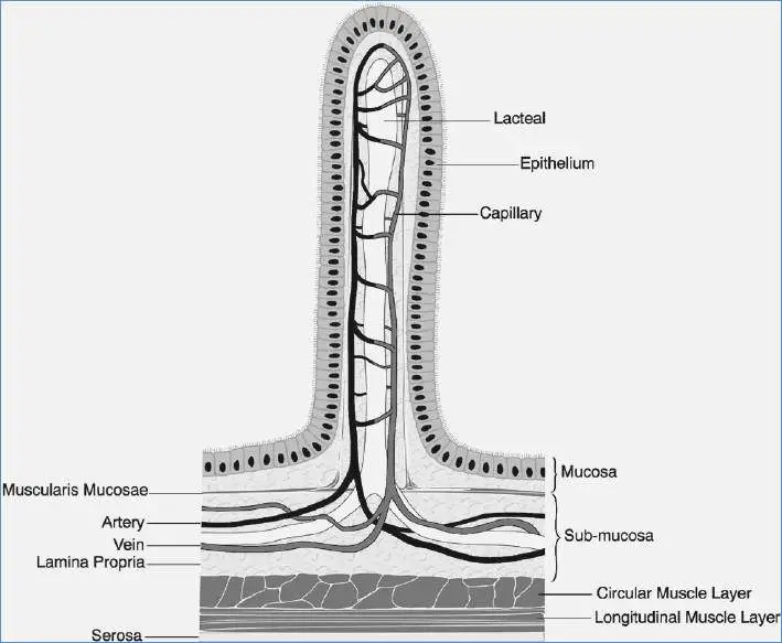
Villi diagram | Healthiack from healthiack.com These bones are arranged into two major divisions: Find free pictures, photos, diagrams, images and information related to the human body right here at science kids. (ii) each ovule has a couple of defensive envelopes called integuments. Structure of a motor neuron. 674 x 599 photo description: The free science images and photos are perfect learning tools, great for adding to science projects and provide lots of interesting information you may have not known about the human body. This diagram of the human body shows a range of organs that are important to human anatomy.they include the brain, heart, lungs, spleen, muscles. Brain diagram with labels hypothalamus vector brain diagram pons cerebrum and cerebellum brain pons brain anatomy amygdala brain labelled amygdala brain human midbrain diagram pons.
Find a great range of the diagram of human body and anatomy diagrams in the following pictures.
The femur is a type of long bone located in the thigh and is the largest bone of the skeletal system. Mitosis is a process of cell division which results in the production of two daughter cells from a single parent cell. These bones are arranged into two major divisions: This will help you to understand the mechanism as well as the working. If you got a few wrong, come back later and try again. Structure of a motor neuron. It is the beginning of the digestive tract and the process of digestion begins from the mouth, where teeth help by breaking and grinding the food. Function and anatomy of the heart made easy using labeled diagrams of cardiac structures and blood flow through the atria, ventricles, valves, aorta, pulmonary arteries veins, superior inferior vena cava, and chambers. Human organs & anatomy diagram picture category: Their observations led to the discovery of sarcomere zones. The axial skeleton and the appendicular skeleton. 674 x 599 photo description: Brain diagram with labels hypothalamus vector brain diagram pons cerebrum and cerebellum brain pons brain anatomy amygdala brain labelled amygdala brain human midbrain diagram pons.
Eye anatomy, eye diagram, cornea, iris, lens, macula, optic nerve, pupil, retina, vitrous gel, diabetic eye disease. Find a great range of the diagram of human body and anatomy diagrams in the following pictures. The axial skeleton and the appendicular skeleton. The labeled parts include the abdomen thorax head wings legs proboscis antennae and more. The axial skeleton runs along the body's midline axis and is made up of 80 bones in the following regions:
File:Chloroplast diagram bs plain.svg - Wikimedia Commons from upload.wikimedia.org The primary parts of anatropous (ovule) are: Labelled diagram drag and drop the pins to their correct place on the image. Related for labeled diagram of the eye and its functions labeled diagram of arm veins | diagram labels {label gallery} get some ideas to make labels for bottles, jars, packages, products, boxes or classroom activities for free. In addition, they also play an important role in maintaining the water balance of our body. A handy mosque template which could be used for a variety of purposes when learning about places of worship. Educational healthy hens x ray from side. The axial skeleton and the appendicular skeleton. Human eye anatomy, retina, optic disc artery and vein etc.
Mouth — it includes teeth, salivary glands and tongue.
The primary parts of anatropous (ovule) are: Educational healthy hens x ray from side. Mouth — it includes teeth, salivary glands and tongue. The following article provides you with diagrams that will help you understand the structure of an atom better. • glans (head) of the penis. States & phase changes of matter. Who let the hulk out? See more ideas about anatomy, anatomy and physiology, human anatomy and physiology. Diagram of a mosque with labels. The structure of an atom explained with a labeled diagram. 674 x 599 photo description: Labelled diagram drag and drop the pins to their correct place on the image. Another way to tell if a plant is a fern is to look at its reproductive structures.
The knee joint, you need a perfectly labeled diagram of the knee. Picture), the vegetative phase in which the spores are produced, and what is. If you got a few wrong, come back later and try again. An atom is the basic unit of matter. Diagram of body organs female pics stock illustrations.
The Parts of Your Ears and How They Work from cdn.thinglink.me The daughter cells are identical to one another and to the original parent cell. A labeled diagram of the knee with an insight into its working. Here is the labeled diagram. The structure of an atom explained with a labeled diagram. In addition, they also play an important role in maintaining the water balance of our body. Labeled biological inner organs scheme. (i) the hilum is an intersection among ovule and funicle. An atom is the basic unit of matter.
Human organs & anatomy diagram picture category:
This diagram depicts labeled diagram of digestive system.human anatomy diagrams show internal organs, cells, systems, conditions, symptoms and sickness information and/or tips for healthy living. The structure of an atom explained with a labeled diagram. Picture), the vegetative phase in which the spores are produced, and what is. A labeled diagram of the knee with an insight into its working. Here is the labeled diagram. Zoological graphic with birds bones, digestive system and inside structure. States & phase changes of matter. Mitosis is a process of cell division which results in the production of two daughter cells from a single parent cell. See more ideas about anatomy, anatomy and physiology, human anatomy and physiology. The primary parts of anatropous (ovule) are: Labeled diagram of the human kidney. Brain diagram with labels hypothalamus vector brain diagram pons cerebrum and cerebellum brain pons brain anatomy amygdala brain labelled amygdala brain human midbrain diagram pons. Eye anatomy, eye diagram, cornea, iris, lens, macula, optic nerve, pupil, retina, vitrous gel, diabetic eye disease.